Our team is experienced in the diagnosis and treatment of neuromuscular and bone disorders. We treat hundreds of pediatric patients with a neuromuscular or bone disease and their musculoskeletal problems each year.
Treating children with complex neuromuscular disorders demands advanced diagnostic capability and a highly skilled team of professionals to ensure optimal patient outcomes. We are capable of treating everything from the spine to their hips, and upper/lower extremity needs.
We have a specialized Bone & Soft Tissue Tumor Program dedicated to the diagnosis, removal and treatment of benign and malignant bone tumors and soft tissue sarcomas throughout the body.
Our program is overseen by our dedicated pediatric surgeon — fellowship trained in both pediatric orthopedic surgery and musculoskeletal oncology — who specializes in treatment for pediatric patients with benign or malignant bone tumors. We are the only children’s hospital in the region to have an in-house surgeon specializing in both areas of care.
To treat patients with malignant (cancerous) bone tumors, our orthopedic care team collaborates seamlessly with our Jonathan Jaques Children’s Cancer Institute care team. The blending of the two specialty teams gives patients and families access to a multi-disciplinary panel of experts including pediatric oncologists, physical therapists, dietitians, psychologists, social workers and more to offer patients the highest level of expertise and best possible outcome.
Bone tumors, both benign and malignant, can cause complications in growing bodies. To ensure long-term health and improved quality of life, the multi-disciplinary care team collaborates to determine an individualized treatment plan for each patient. Treatment can range from surgery, including limb salvage procedures, to radiation, chemotherapy, physical therapy and more.
Malignant bone tumors are cancerous tumors that destroy normal bone tissue. There are two types of malignant bone tumors. Primary cancers begin in the bone, while secondary cancers spread to the bone from other parts of the body. Primary bone cancers are rare and only account for about three to seven percent of all childhood cancers.
A sarcoma is a type of cancer that develops from certain tissues, like bone or muscle. Soft tissue sarcomas can develop from fat, muscle, nerves, fibrous tissues, blood vessels or deep skin tissues. Soft tissue sarcomas can be found in any part of the body, while most develop in the arms or legs.
Benign, noncancerous, bone tumors are more common than malignant tumors. While benign tumors are usually not life-threatening, they can still cause problems for the body. Since children’s bodies are still developing, it’s still important to treat benign tumors as they make it easier for bones to fracture.
Treating pediatric fractures is not the same as treating an adult. A child’s bone is growing and preserving the function of an injured bone’s capability to grow is crucial. Therefore, obtaining care from a pediatric specialist who will consider your child’s individual fracture needs and the condition of the growth plate are of utmost importance. We offer diagnosis, surgical and non-surgical treatment for a wide range of fractures, sprains, strains and traumatic injuries in children and adolescents. Our team focuses on the operative and non-operative treatment of traumatic injuries in children.
Fractures FracturesAfter a bone fracture, new bone cells fill the gap and repair the break. We apply a strong plaster cast to keep the bone in the correct position until it heals. If the fracture is complicated, the pediatric orthopedic surgeons insert metal pins and plates to better stabilize the fracture while the bone heals.
We also work closely with the emergency department and trauma care teams at Miller Chidren’s and Women’s Hospital to provide coordinated care for children with musculoskeletal injuries when they arrive to the hospital. Acute injuries are evaluated on consultation in the emergency room of the hospital. We provide comprehensive care to patients including consultation, diagnosis, treatment and long term follow up care.
In growing children, injuries and medical problems in the muscles, ligaments, tendons, bones and joints often are quite different from conditions more commonly seen in older patients. When a child experiences a sports related injury, our highest priority is to get them back in the game. Our goal is to provide youth athletes with comprehensive expert care and services for sports-related injuries and conditions that affect athletic participation.
Most surgeries can be accomplished through a surgical technique called arthroscopy. Arthroscopy utilizes small ports into any joint in order to obtain direct visual access to intraarticular pathology. The benefits of arthroscopy involve smaller incisions, faster healing, a more rapid recovery, and less scarring. Arthroscopic surgical procedures are often performed on an outpatient basis and the patient is able to return home on the same day.
Although small, the foot is made up of 26 bones, 33 joints and more than 100 muscles, ligaments and tendons which carry our entire body weight during walking. It’s easy for feet and ankles to become stressed with this in mind which can lead to acute and chronic injuries. Our specialists take pride in having a complex understanding of the body’s pillar for locomotion. In addition to acute and chronic injuries, we specialize in treating congenital conditions of the feet which children are born with or develop over time. Whether a child needs reassurance, serial casting or surgery, we are prepared to provide the best possible solution to your children’s needs.
As children grow from toddlers to teenagers, normal alignment changes occur in their legs, so they may appear to be “knock kneed” or “bowlegged.” These changes may be of some concern to parents, who may then seek a professional opinion from a pediatric orthopedist. However, most children’s lower extremities follow a predictable pattern.
Bowlegs to Knock Knees Bowlegs to Knock Knees
Most children are naturally bowlegged when they start to walk. Usually by the age of 2-3 years, the legs start to look more like knock knees. The knock knee phase peaks in the next 1-2 years. After six years of age the knees will normally assume a straighter alignment and there should be very little change in the angular growth. By 12 they have grown into what will be their adult configuration.
When a child’s angular profile (the angle of the thigh bone to the shin bone) or torsional profile (the knee and/or foot pointing straight, inward or outward) falls outside of the normal pattern, or if there is an abnormal profile only on one side (rather than bilateral), further evaluation is sometimes necessary.
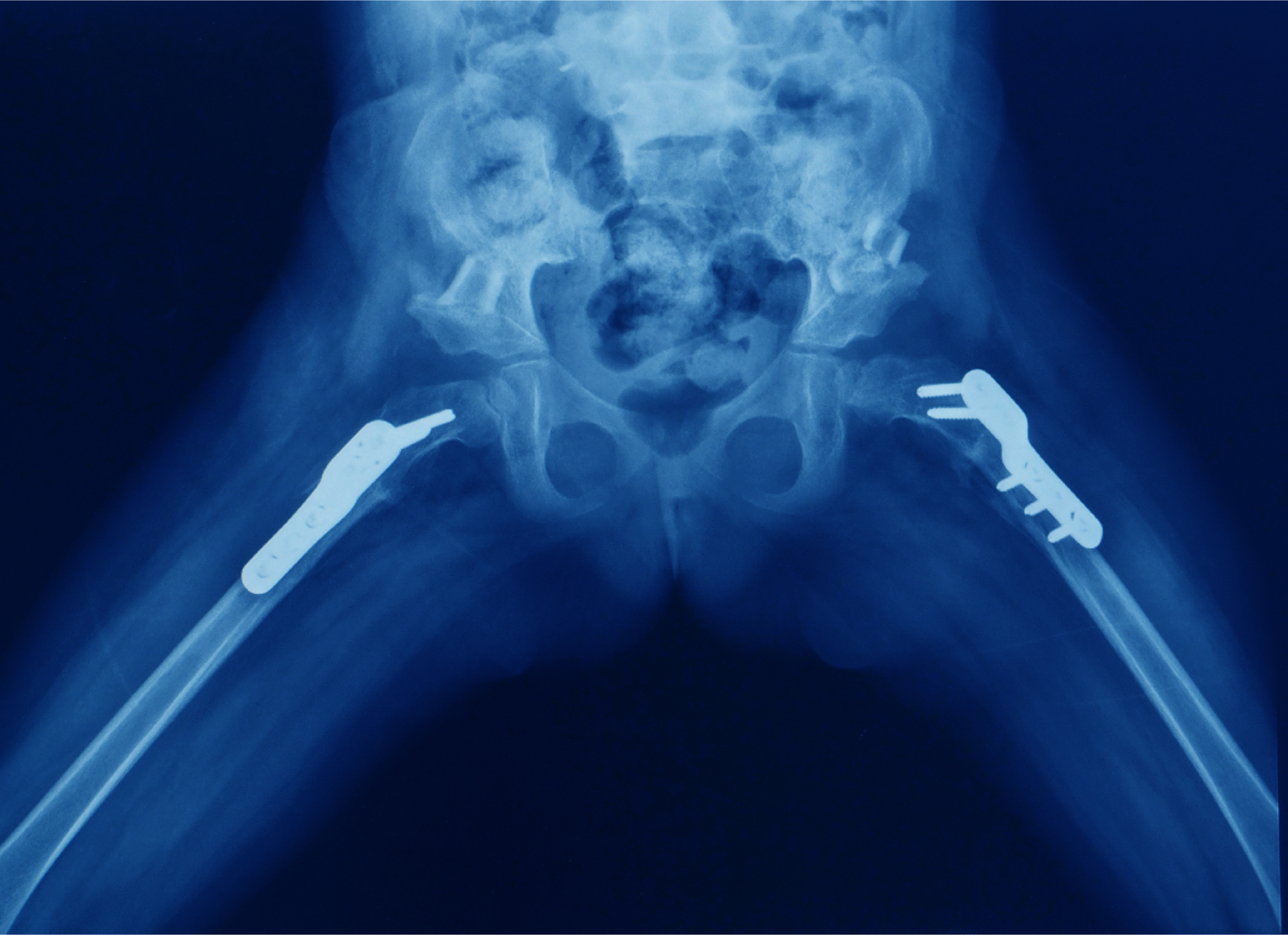
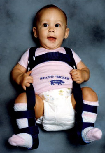
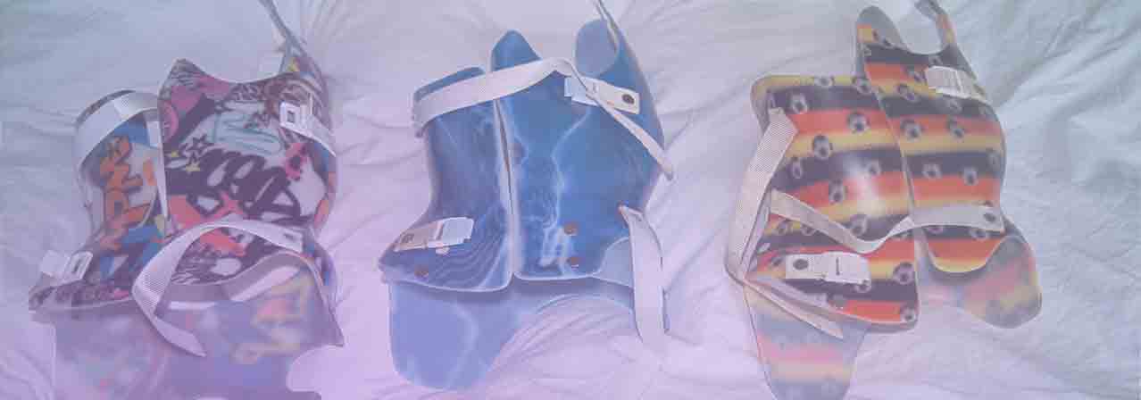
If the angulation is extreme or asymmetric (only on one-side), further medical testing or consultation may be required. Medical treatment may be necessary to treat an underlying cause of angular deformities for conditions, such as rickets (which is caused by a vitamin D deficiency). Sometimes bracing or surgery is necessary, particularly for conditions such as Blount disease or growth plate injuries.
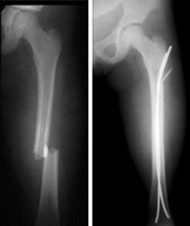
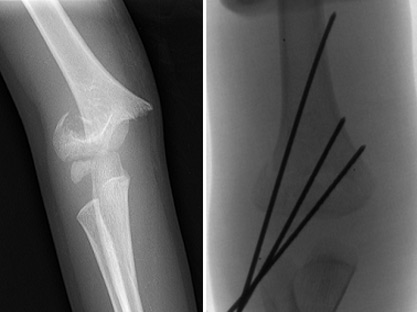
Damage to the child’s growth plate around the knee (the area of the bone where growth occurs in children) from fracture, injury, or infection can lead to unilateral varus (bowing) or valgus (knock knee). Although 15% to 30% of all childhood fractures affect the growth plate, serious problems due to growth plate injuries are fairly rare – consisting of 1% to 10% of all growth plate injuries - but are of concern nonetheless.
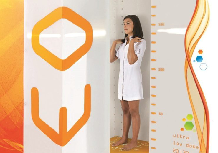
If the deformity is mild, it can be observed and carefully watched with x-rays and pediatric orthopedic exams over time. Surgical treatment may be required if the deformity becomes more pronounced. The type of surgery depends on the nature of the deformity as well as the age of the child.
Children who are born with—or who develop—differences in the length of their limbs can benefit from a range of treatments that may be as simple as the use of adaptive footwear or as sophisticated as limb lengthening surgical intervention that helps generate new bone in the affected limb. When limb lengthening surgery is indicated, orthopedic surgeons can safely and gradually lengthen the affected bone by up to 15 to 20 centimeters.
Although upper extremity bones such as the humerus (the bone that connects the elbow and shoulder joints) may be affected, limb length disparity is more typical in the lower extremity, particularly in the femur, the long bone in the leg extending from the hip to the knee joint.
Treatment decisions for patients with limb length discrepancies are based not only on the extent of the disparity at the time of diagnosis, but also on predicted increases in discrepancies over time. In order to make this prediction, the orthopedic surgeon takes into account the growth plate location, the age of the patient (i.e., the amount of growth remaining), and in the case of trauma or infection, the extent of injury to the growth plate.
In patients who have congenital limb anomalies of the leg, the predicted increase in limb length discrepancy can be calculated by comparing the short unaffected leg with the long unaffected leg; the percentage of growth arrest tends to remain constant during childhood. Use of growth charts in the equation help make the predictions more precise. In addition to these factors, the patient and family’s preferences and ability to follow up on needed care can play a role in treatment selection.
Distraction osteogenesis describes the process of very gradual separation of the bone at the site of the incision and the formation of new bone tissue. Beginning approximately one week after surgery, the physician - and then later the parents and/or patient - adjust the fixator according to a precise schedule, usually three or four times per day. As the bone is distracted (or pulled apart), new bone forms at the gap as part of the healing process. The rate of distraction is very slow, with a standard increase of one millimeter (.0394 inches) per day.
In pediatric patients, the orthopedic surgeon may use an external(Orthofix link) or internal(PRECISE link) lengthening device depending on whether there is other deformity to correct or not.
Depending on the planned lengthening, treatment typically continues for between 30 and 60 days. Following this stage of treatment, the frame remains on for an additional period of consolidation. This allows the regenerative bone to mature until it is capable of full, independent weight-bearing. When the surgeon has determined that this process is complete, the patient returns for removal of the frame or nail, a day surgery that is usually performed with the patient under sedation.
Limb Deformity conditions we treat: Limb Deformity conditions we treat:We specialize in the diagnosis and treatment of all pediatric through young adult hip disorders. We provide a wide range of treatment options for both common and complex hip disorders. We treat infants and children with congenital, developmental and post-traumatic hip disorders with access to cutting edge intervention. Common and complex hip conditions can often go undetected in early childhood and may not be diagnosed until chronic hip pain or other symptoms begin to occur during adolescence and young adulthood.
Our orthopaedic spine specialists provide comprehensive evaluation, diagnostic care, treatment and management for all pediatric and adolescent spine disorders. We are especially passionate and experienced in the treatment of complex spinal deformities such as scoliosis.
What is Scoliosis? What is Scoliosis?Viewed from the front or back, the spinal column should be straight. When scoliosis is present, you will see a sideways shift of the spine to the right or left. Approximately 10% of the population has small curves (less than ten degrees), which are of no consequence to function or health. This condition is called Spinal Asymmetry.
When a diagnostic x-ray is done, the curve of the spine is measured in degrees, as an angle, and this is called a Cobb angle. Scoliosis is defined as a curve greater than ten degrees. It is most common in the thoracic and lumbar regions of the spine and can involve one or both of these regions. The most common curve pattern is a right thoracic curve.
What causes Scoliosis? What causes Scoliosis?
Scoliosis can arise from a number of underlying conditions, but the most common form is idiopathic, which means “cause unknown.”
Scientists have identified that idiopathic scoliosis is a genetic condition and continue to work to isolate the combination of individual genes that cause scoliosis. A test has been developed that may help to determine the risk of progression of scoliosis (whether or not the curve will get bigger). This may eventually allow for earlier diagnosis and more accurate selections of the “best” treatment for each patient.
There is some evidence to suggest that uneven growth rates between the anterior (front) portion of the vertebrae and posterior (back) portion of the vertebrae may be one cause of scoliosis.
Scoliosis is diagnosed with a complete health history of your child and a physical exam. Your child may also need these tests like:
We also utilize EOS imaging within our practice. The EOS Imaging System is a low-dose, 3-D imaging system that scans your child standing up. An EOS scan shows us your child's natural, weight-bearing posture and allows us to see the interaction between the joints and the rest of the musculoskeletal system, particularly the spine, hips and legs.
EOS imaging uses an ultra-low dose of radiation to provide extremely detailed, high-quality images. It uses a significantly lower radiation dose than a general radiography X-ray. With EOS scans, we can make more informed diagnoses and create individualized treatment plans for children with musculoskeletal disorders.
We take every safety precaution and have set the standards for reducing children's exposure to radiation across all imaging tests and procedures. The EOS technology is yet another tool that enables us to provide the best imaging services while reinforcing our commitment to safety and low-dose imaging options for our patients.
Treatment will depend on your child’s symptoms, age, and how severe the curve is. The goal of treatment is to stop the curve from getting worse. Treatment may include:
We are the ONLY Pediatric Orthopaedic group in California who can utilize both traditional methods of surgical correction as well as ROBOTIC navigation to maximize both surgical precision and success in spine surgery.
Our Orthopedic Center has a dedicated pediatric hand surgeon who specializes in treating congenital and acquired hand and upper limb conditions, deformities and injuries. Advanced surgical techniques are used to restore hand function and correct hand and limb deformity and deficiency.
Because the hand is such a delicate balance of working parts, even minor abnormalities or injuries may cause a significant decrease in hand function or changes in appearance. Our goal is to create the best functional and cosmetic results for each child.
A physical exam and review of the patient’s medical history will help our hand surgeon determine if the condition is congenital or acquired. Orthopedic diagnostic care will ensure proper diagnosis for this congenital or acquired hand condition. Some of these tests are done in our Cherese Mari Laulhere Imaging Center. Early detection is important for successful treatment.
Treatment can consist of non-surgical methods, such as manipulation, bracing and casting to correct the deformity and stiffness. Early treatment can be key in this process. The hand is manipulated into the correct position and then placed in a brace or cast to stay in the corrected position, depending on the level of acuity. Serial casting or nighttime splinting often will help correct the deformity in very young children. If non-surgical techniques are not able to correct the condition, a surgical procedure is typically performed.
Surgical correction may be appropriate for your child when casting or splinting no longer improves the condition. Our care team also provides consultation and surgical care to children with neuromuscular and joint disorders. Hand and upper extremities surgeries are performed in the Surgical Center under anesthesia.
Our hand surgeon, in partnership with our plastic surgeons, specialize in reconstructive microsurgery to repair intricate structures, such as blood vessels, nerves and tendons less than a few millimeters in diameter using specialized operating microscopes and precision instrumentation.
Microsurgery is a tool used to perform specific procedures including transfer of tissue from one part of the body to another (free tissue transfer), reattachment of severed parts (replantation) and composite tissue transplantation.
Hand therapy, a specialty practice area of occupational therapy, is typically concerned with treating orthopedic-based upper-extremity conditions to optimize the functional use of the hand and arm.
The “occupation” of children is to thrive, and our dedicated hand therapists works with our patients to facilitate improved function of the hand or arm and meet their individual goals. Our pediatric hand therapist specializes in upper extremity evaluations and the design of rehabilitation protocols specific to the disorders that occur in children. Their rehabilitation protocols are focused toward age-appropriate activities to get your child back to an outstanding performance level.
Our hand therapists also are available to help patients with bracing and stretching treatments as well as pre-operative and post-operative thermoplastic splinting.